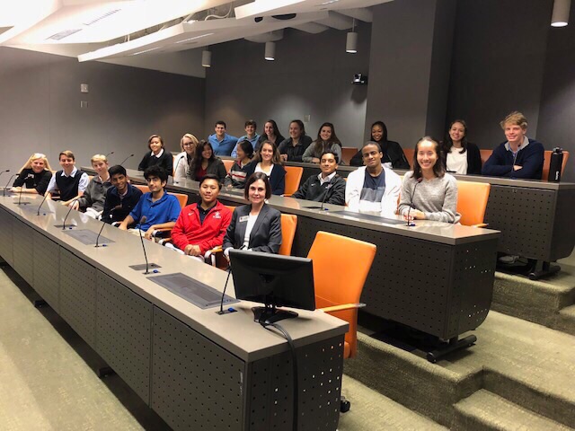
Anatomy and Physiology students travel to Methodist Hospital, view robotic-assisted surgery
Used with permission from Lizo McNeely.
The students met with several doctors, who spoke to the class about their training and work.
October 26, 2018
On Oct. 17, Anatomy and Physiology students traveled to Methodist Hospital for their annual fall field trip. The students received tours of several labs — led by different doctors who spoke to them about their background and training — and watched a live-stream of a robotic-assisted surgery in one of Methodist’s new viewing rooms.
According to Anatomy teacher Paula Angus, the purpose of the field trip was for her students to see how the extensive material they cover in class is applied in the real world.
“Part of our tour involved going through the blood center,” Angus said. “We saw where blood is kept and sorted, and [the students] got a better understanding of why blood type has to be carefully separated — to ensure that they are using the right blood type in an emergency.”
The students also went on a tour with two prominent pathologists who showed them different areas of the pathology lab and slides taken from various body parts, such as an eyeball, ear and breast tissue.
“That tour was really interesting and the doctors were very insightful,” senior Elizabeth McNeely said. “Seeing the slides under the microscope was cool because it was giving us more context as to what it would be like looking at these things if we were ever to become pathologists.”
The class also met with two neurosurgeons. They examined and identified different sections of a real human brain, as well as a spinal cord. This part of the tour was a great opportunity for some of the students to gain insight into a potential career path.
“A lot of us in this class want to be doctors, but some are really interested in certain specialties, like neurosurgery,” McNeely said. “This field trip was a great opportunity because they got to speak with the neurosurgeons about their expertise and gain some perspective about their field.”
The highlight of the trip for many students was a surgery viewed from a closed-circuit television in a viewing room. The surgery was a new procedure: a robotic-assisted repair of a hiatal hernia.
“A hiatal hernia occurs when the upper part of the stomach pushes up through the diaphragm,” Angus said. “They have to pull the stomach down and add an artificial valve that will prevent a hernia from recurring. We never know which surgery we’re going to get to watch, so it’s always a fun surprise finding out.”
While she greatly enjoyed watching the surgery and listening to the doctor’s accompanying comments, McNeely remarked on its gory details.
“The surgery was not for the faint-hearted,” McNeely said. “At one point, the surgeons accidentally cut a blood vessel, and blood just came spurting out everywhere. It was absolutely incredible to watch, but a bit gruesome.”
The trip was a success, according to Angus, and “a good preparation” for the class’ spring field trip to the Texas Orthopedic Hospital, where students will watch a limb replacement in a surgical room.
“You can learn so many things in the classroom, but having that hands-on experience is just amazing,” McNeely said. “We’re going off to college next year, and already knowing what the environment in a hospital helps with important career decisions.”